
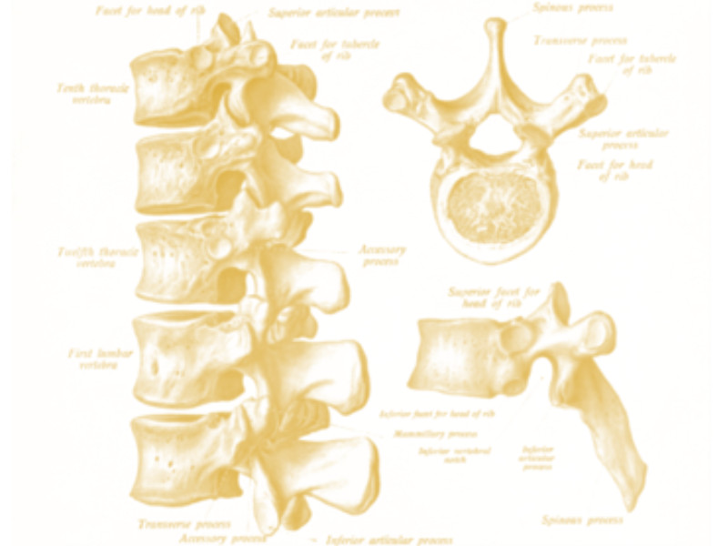
Lumbar Spine Surgery
You are having surgery on your lumbar spine, which is the lower segment of your vertebral column consisting of five individual vertebrae. The spinal cord ends at the first lumbar vertebra and the spinal nerves continue to descend the rest of the length of the vertebral column. This bundle of spinal nerves are called the cauda equina which is Latin for horse’s tail owing to their resemblance to the same.The nerves exit the vertebral column at the appropriate level for them to innervate the lower parts of the body. Their precise exit point puts them at risk of injury during lumbar spine surgery.
Monitoring
In order to protect the spinal nerves, an NMA neurophysiologist will be monitoring your nervous system during the entirety of your procedure. Neuromonitoring consists of a variety of testing modalities to ensure that signals are moving up and down the nervous system correctly during surgery. If there is a change in neurological function detected, the neurophysiologist will work with the surgeon to identify the problem and correct it, avoiding a bad surgical outcome.During lumbar surgery, the spinal nerves and spinal cord are protected by two main tests: somatosensory evoked potentials (SSEPs) and electromyography (EMG). The SSEP test is conducted by stimulating nerves in your ankles and wrists and tracing the nerve signal from the point of stimulation all the way to the brain. If there is a potential injury to the spinal nerves or spinal cord, we will see an interruption of this signal and inform the surgeon. EMG testing is a direct way of monitoring the health of the spinal nerve. If the spinal nerves are injured or irritated the muscles that they innervate will fire and contract. We can measure that contraction in the form of an electrical potential. Another way that EMG is used during a lumbar fusion surgery is to test the positioning of the pedicle screw that is misguided and can potentially injure the nerve. To help avoid that outcome, the neurophysiologist can stimulate the screw electrically and if it is placed near the nerve root, a muscle potential will be recorded. This allows the surgeon to go back and redirect the position of the screw.