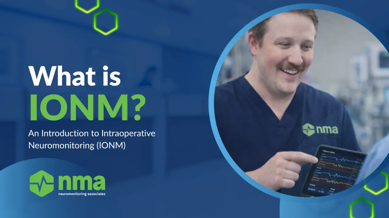
What Is Intraoperative Neuromonitoring (IONM)? Techniques, Applications, and Clinical Impact
In today’s high-stakes neurosurgical environment, precision is everything. As procedures become more complex and patient expectations rise, safeguarding neurological function has never been more critical.
Intraoperative neuromonitoring (IONM) has emerged as a cornerstone of modern neurosurgery—offering real-time insight into the functional integrity of neural pathways and empowering surgical teams to make data-driven decisions in the moment.¹
But what exactly is IONM, and how does it impact surgical outcomes?
What Is IONM and Why Does It Matter?
IONM refers to the continuous, real-time monitoring of the nervous system during surgery, enabling the detection of functional changes before they result in permanent injury.¹ By providing both anatomical and functional feedback, IONM allows surgical teams to intervene immediately when neural structures are at risk—significantly improving patient safety and postoperative outcomes.¹
IONM is not a single technique, but a suite of complementary modalities designed to monitor specific neural pathways.
Core IONM Modalities
1. Somatosensory Evoked Potentials (SSEPs) SSEPs monitor sensory pathways, with changes in amplitude or latency indicating potential compromise to neural transmission.¹
2. Electromyography (EMG) EMG evaluates motor nerve integrity by recording muscle responses to nerve stimulation, providing early warning signs of nerve irritation or injury.¹
3. Motor Evoked Potentials (MEPs) and Direct Cortical Stimulation (DCS) MEPs assess motor pathway integrity, while DCS allows precise cortical mapping—both critical for preserving motor function during high-risk procedures.¹
4. Brainstem Auditory Evoked Potentials (BAEPs) BAEPs monitor auditory pathways and are particularly valuable in posterior fossa and brainstem-related surgeries.¹
5. Electrocorticography (ECoG) and SEEG ECoG enables direct cortical recording for high-resolution mapping of motor, sensory, and language areas, making it essential for identifying eloquent cortex and epileptic activity.¹ SEEG extends this capability deeper, allowing precise localization of epileptogenic zones and functional regions in complex epilepsy cases.¹
6. Visual Evoked Potentials (VEPs) VEPs evaluate optic pathway function, helping protect visual outcomes during surgeries near visual structures.¹
Together, these techniques form a multimodal monitoring strategy, improving diagnostic accuracy and reducing the limitations of single-modality monitoring.¹
Clinical Applications of IONM
The true value of IONM lies in how it is applied. Across neurosurgery, it serves as a real-time safeguard—guiding intraoperative decisions and protecting patient function.
1. Intracranial Tumor Resection: Balancing Resection with Preservation
In brain tumor surgery, the goal is to maximize tumor removal while preserving neurological function. IONM supports this by enabling continuous mapping and monitoring of eloquent cortex and subcortical pathways throughout the procedure.¹
Techniques such as dynamic subcortical mapping, MEPs, SSEPs, ECoG, and DCS help identify critical regions, detect early functional changes, and protect motor eloquent areas, allowing surgeons to adjust in real time.¹ This transforms tumor resection into a dynamic, function-guided process rather than a purely anatomical one.
2. Neurovascular Surgery: Detecting Ischemia Before It’s Irreversible
Neurovascular procedures carry a high risk of cerebral ischemia. IONM provides an early warning system by identifying functional changes caused by reduced blood flow before permanent damage occurs.¹
By using modalities such as SSEPs, MEPs, EEG, and ECoG, surgical teams can detect ischemic trends, intervene immediately, and reduce the risk of stroke or long-term deficits.¹
3. Epilepsy Surgery: Precision Mapping for Better Seizure Outcomes
For patients with drug-resistant epilepsy, surgical intervention requires precise localization of seizure-generating regions. IONM techniques—particularly ECoG and SEEG—allow clinicians to identify epileptogenic zones while preserving surrounding functional cortex.¹
In combination with direct cortical stimulation, IONM supports accurate mapping of motor, sensory, and language areas, improving surgical precision and long-term seizure outcomes.¹
4. Spinal Surgery: Protecting the Spinal Cord in High-Risk Procedures
Spinal surgery remains one of the most established applications of IONM due to the risk of neurological injury. Techniques such as MEPs, SSEPs, and D-waves continuously monitor spinal cord integrity during instrumentation and correction.¹
Multimodal monitoring enhances reliability, allowing early detection of mechanical or ischemic compromise and enabling timely surgical adjustments to prevent permanent deficits.¹
5. Peripheral Nerve Surgery: Real-Time Nerve Identification and Preservation
In peripheral nerve procedures, IONM provides critical insight into nerve function and integrity. Using EMG and nerve action potentials, surgeons can differentiate between healthy and damaged tissue during intervention.¹
This real-time feedback supports more accurate nerve repair, reduces uncertainty, and improves functional recovery in trauma, decompression, and tumor-related cases.¹
6. Cross-Specialty Impact: One Framework, Multiple Applications
Across all neurosurgical settings, IONM provides actionable data that directly informs intraoperative decisions.¹ It allows clinicians to detect early neural compromise, intervene immediately, and preserve function while advancing surgical goals.
Clinical Considerations: Why Execution Matters
While IONM is powerful, its effectiveness depends on proper execution. Factors such as anesthetic management, physiological stability, and technical setup can significantly influence signal accuracy.¹
Stable anesthetic delivery—often via total intravenous anesthesia (TIVA)—is essential for reliable monitoring, while variables like hemodynamic changes or hypothermia can distort signals.¹ Multidisciplinary coordination ensures accurate interpretation and appropriate response to intraoperative changes.¹
IONM, therefore, is not just a tool—it is a clinical system requiring expertise, consistency, and collaboration.
Key Takeaways
- IONM provides real-time functional monitoring that improves surgical safety and outcomes across neurosurgery¹
- It supports critical applications in brain tumor, vascular, epilepsy, spinal, and peripheral nerve procedures¹
- Multimodal monitoring enhances reliability and reduces the limitations of single techniques¹
- IONM enables earlier detection of neural compromise and immediate intraoperative intervention¹
- Its success depends on proper anesthetic management, technical execution, and team coordination¹
- IONM is best understood as a clinical decision-making framework—not just a technology¹
Article Citation
Guzzi, G.; Ricciuti, R.A.; Della Torre, A.; Lo Turco, E.; Lavano, A.; Longhini, F.; La Torre, D. Intraoperative Neurophysiological Monitoring in Neurosurgery. Journal of Clinical Medicine. 2024;13:2966. https://doi.org/10.3390/jcm13102966